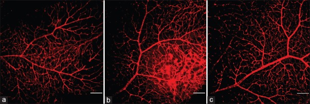
Figure 3.
Characterization of the retinal vasculature in the nondiabetic control and STZ-induced diabetic mice. Freshly dissected retinas were immediately fixed and stained with an endothelial cell marker and visualized. Nondiabetic mice had an organized retinal vascular branching pattern (a). After 18 weeks of diabetes, the vascular distribution in the retinas was dense and disorganized (b), but this improved after LIF treatment (c) (bar: 100 μm). STZ: Streptozotocin; LIF: Leukemia inhibitory factor.

