Figure 1.

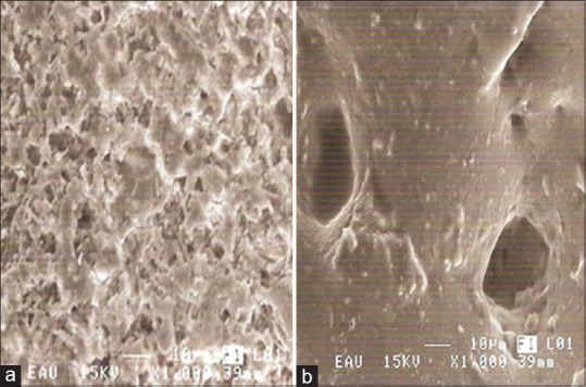
Scanning electron microscopy observation of the Ivoclar porcelain after surface treatments at ×1000 and bar marker indicating 10 μm. (a) Surface with etching hydrofluoric acid (9.5%). (b) Surface with air abraded with aluminum oxide

Scanning electron microscopy observation of the Ivoclar porcelain after surface treatments at ×1000 and bar marker indicating 10 μm. (a) Surface with etching hydrofluoric acid (9.5%). (b) Surface with air abraded with aluminum oxide