Figure 3.

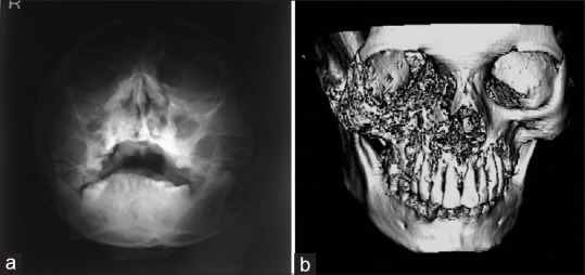
(a) Paranasal sinus radiograph showing haziness of the right maxillary sinus with destruction of sinus walls. (b) Three-dimensional computed tomography scan showing hyperdensity of maxillary antrum with destruction of all boundaries

(a) Paranasal sinus radiograph showing haziness of the right maxillary sinus with destruction of sinus walls. (b) Three-dimensional computed tomography scan showing hyperdensity of maxillary antrum with destruction of all boundaries