Fig. 1.

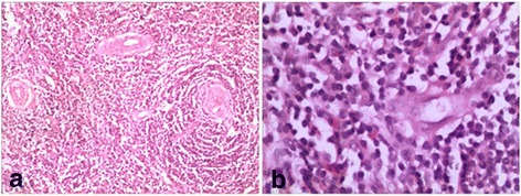
a histology of the tissue showed proliferation of lymphoid follicles (H&E, 100X); (b), lymph node with lymphoid cells in an “onion skin” pattern with a hyaline center (H&E, 100X)

a histology of the tissue showed proliferation of lymphoid follicles (H&E, 100X); (b), lymph node with lymphoid cells in an “onion skin” pattern with a hyaline center (H&E, 100X)