Figure 1.
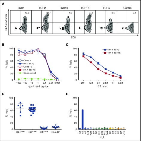
HA-1 TCR–transduced CD8+T cells kill HA1+target cells. (A) Flow cytometry showing HLA-A2/HA-1 multimer staining of CD8+ T cells transduced with HA-1 TCR LV (TCR 1, 2, 10, 16, 5) and CD8+ T cells transduced with TCRs specific for a different minor H antigen (control). (B-E) CRAs to evaluate specific lytic activity. (B) Lysis of T2 target cells pulsed with HA-1 peptide at various concentrations by TCR-transduced CD8+ T cells (solid lines and symbols; TCR2, blue circles; TCR16, red squares), HA-1–specific T-cell clones (dashed lines, open symbols: clone 2, blue circles; clone 16, red squares), or T-cell clone control (diamonds). (C) Lysis of HLA-A2+HA-1+ LCL by CD8+ T cells transduced with HA-1 TCR2 (circles) or HA-1 TCR16 (squares) at various E:T ratios. (D) Lysis of HLA-A2+/HA-1+ homozygous (H/H) (circles, n = 7), HLA-A2+/HA1+ heterozygous (H/R) (squares, n = 22), HLA-A2+/HA-1– (R/R) (triangles, n = 17), or HLA-A2–negative (inverted triangles, n = 41) hematopoietic cell (LCL) targets by HA-1 TCR2–transduced CD8+ T cells. (E) Lysis of LCL with common HLA alleles by HA-1 TCR2–transduced CD8+ T cells. *An E:T ratio of 20:1 was used unless otherwise specified. Data comparable with that shown in panels D and E were also obtained with HA-1 TCR16 (data not shown).

