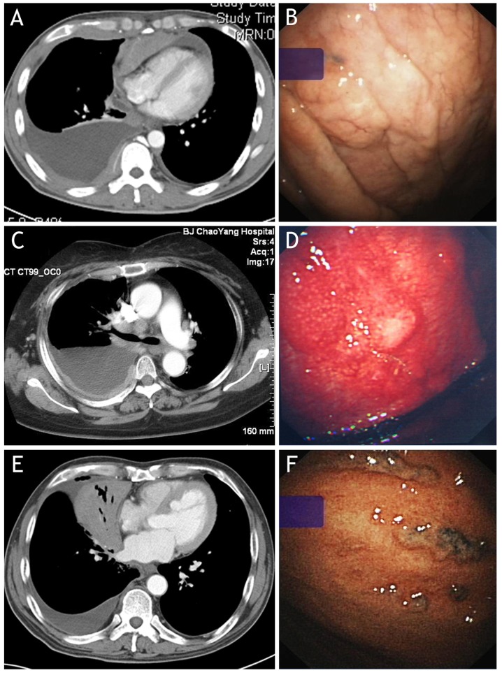
Figure 1.
Abnormalities observed on contrast-enhanced CT (left panels) and under medical thoracoscopy (right panels) in patients with malignant pleural effusion induced by non-Hodgkin's lymphoma. (A) CT reveals diffuse pleural thickening and pleural nodules in the right hemithorax; (B) thoracoscopy of the same patient of (A) reveals diffuse pleural thickening on parietal pleura. (C) CT reveals diffuse pleural thickening and nodules in the right hemithorax; (D) thoracoscopy of the same patient with (C) reveals diffuse pleural thickening, scattered nodules and hyperemia on the parietal pleura. (E) CT reveals isolated pleural effusion in right hemithorax and lung atelectasis of the right middle lobe, with no discernible pleural abnormalities; (F) thoracoscopy of the same patient with (E) reveals small nodules with an irregular distribution on the parietal pleura. CT, computed tomography.

