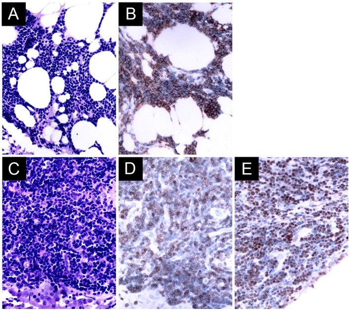
Figure 2.

(A) Pleural biopsy from a patient with non-Hodgkin's extranodal marginal zone B-cell lymphoma reveals a diffused small cell neoplasm, with slightly irregular nuclei with moderately dispersed chromatin non-conspicuous nucleoli by H&E staining. (B) Immunohistochemistry reveals that the neoplastic cells were CD20-positive. (C) Pleural biopsy from a patient with non-Hodgkin's T-lymphoblastic lymphoma showing neoplastic cells of a small size with a high nuclear-to-cytoplasmic ratio and with condensed nuclear chromatin and no evident nucleoli by H&E staining. Immunohistochemistry shows that the neoplastic cells were (D) CD3-positive, and (E) terminal deoxynucleotidyl transferase-positive. Original magnification, ×400 for all panels. H&E, hematoxylin and eosin; CD20, cluster of differentiation 20.
