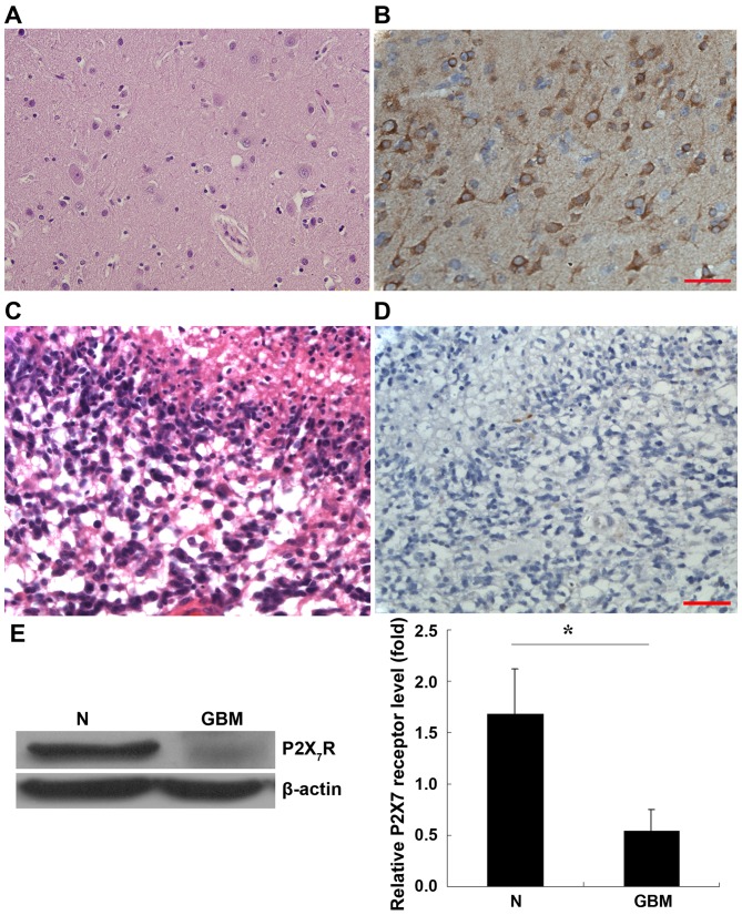
Figure 1.
Expression of P2X7R in GBM and normal brain tissue. P2X7R expression was determined by immunohistochemistry staining. (A) H&E staining of normal brain tissue. (B) Normal brain tissue stained for P2X7R expression. (C) H&E staining of GBM. (D) GBM stained for anti-P2X7R expression. (E) Western blot analysis. P2X7R protein was significantly reduced in human GBM compared with the peripheral normal brain tissue. Data are expressed as the mean ± standard error of mean. *P<0.05. Scale bar, 50 µm. P2X7R, p2X purinoceptor; GBM, glioblastoma; H&E, hematoxylin and eosin.

