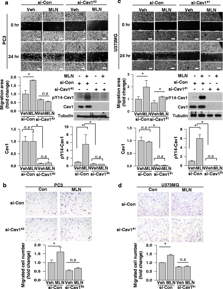
Fig. 3.

Phosphorylated caveolin-1 is in charge of MLN4924-induced cell migration. a, c Scratch-based wound healing assays were performed for 24 h in PC3 (a) and U373MG (c) cells which were depleted of caveolin-1 using siRNAs (#2 and #1, respectively) and si-control in the presence of MLN4924 (0.25 μM and 0.5 μM) or DMSO (top). The migration areas were calculated using ImageJ (middle, left). Proteins in cells lysates were analyzed by Western blotting (middle, right). The efficiency of the Caveolin-1 knockdown and magnitude of the phosphorylation of Caveolin-1 was quantified based upon the relative level of β-tubulin (bottom). b, d Transwell migration assays were performed in PC3 (b) and U373MG (d) cells which were depleted of caveolin-1 using siRNAs (#2 and #1, respectively) and si-control in the presence of MLN4924 (0.25 μM and 0.5μM) or DMSO for 24 h (top), and migrated cells were counted (bottom). Each bar represents the means + standard deviation of results from three independent experiments. * denotes P < 0.05 and n, s, does P > 0.05 between the indicated groups. Scale bar = 200 μm
