Fig. 1.

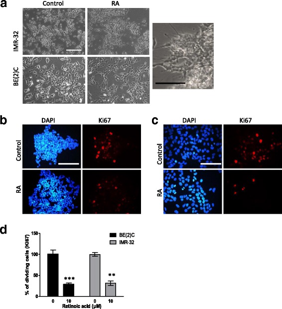
ATRA promotes a reduction in cell proliferation and change in morphology in IMR-32 and BE(2)C cells. a Morphological changes in IMR-32 and BE(2)C after 3 days of treatment with 10 μM ATRA (RA), an enlarged view of IMR-32 cells is displayed showing a number of cellular extensions of variable length. b DAPI stained (blue) and Ki67 stained (red) BE(2)C cells following three days of treatment with 10 μM ATRA or DMSO (control). c DAPI stained (blue) and Ki67 stained (red) IMR-32 cells following three days of treatment with 10 μM ATRA or DMSO. d Graph to show the reduction in cell proliferation following treatment with ATRA. Each bar represents three biological replicates and at least 9 fields per experiment. ** p < 0.01 and ***p < 0.001. Error bars are standard error (SE). Scale bar is 100 μM
