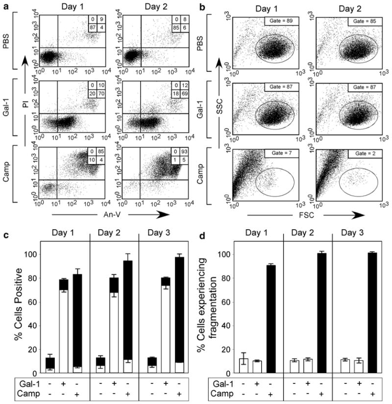
Fig. 6.
Gal-1 induces continuous PS exposure in the absence of cellular fragmentation. (a) Cells were incubated with PBS, 10 μM iodoacetamide-alkylated Gal-1 (iGal-1), or 10 μM Camp for 1 or 2 d as indicated, followed by detection for PS exposure by An-V-FITC staining and PI exclusion. (b) Cells were incubated with PBS, 10 μM iGal-1, or 10 μM Camp for 1 or 2 d as indicated, followed by examination for cellular fragmentation as indicated by changes in forward (FSC) and side scatter (SSC) profiles of cells. Gate = % of cells experiencing no fragmentation. (c ) Quantification of cells treated in (a). White bars = % An-V+, PI−; black bars = % An-V+, PI+. (d) Quantification of cells treated in (b). This research was originally published in Molecular Biology of the Cell. Stowell SR, Karmakar S, Arthur CM, Ju T, Rodrigues LC, Riul TB, Dias-Baruffi M, Miner J, McEver RP, Cummings RD. Galectin-1 induces reversible phosphatidylserine exposure at the plasma membrane. 2009 Mar;20(5):1408–18

