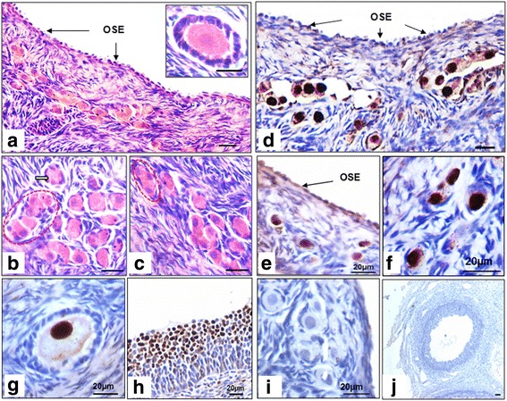
Fig. 11.

PCNA immuno-localization on sheep ovarian sections. H&E staining of ovarian sections (a) shows a distinct layer of OSE cells (b-c) cohort of cytoplasmically connected oocytes i.e. a germ cell nest surrounded by few pre-granulosa cells. These structures are referred to as ovigerous cords in literature (Smith et al. 2014). Few primordial and primary follicles were located in the cortical region whereas large oocytes were present in medulla region. d-h Immuno-localization with PCNA showed few OSE cells positive for PCNA, cluster of oocytes/germ cell cyst, individual primordial and primary follicles located in cortical region of ovary showed strong nuclear PCNA. Interestingly surrounding granulosa cells and stromal cells were completely negative for PCNA. Large Graffian follicle in medulla region showed weak PCNA expression in both nucleus and ooplasm of oocytes however, (h) surrounding granulosa cells were strongly positive for PCNA. i & j Negative control
