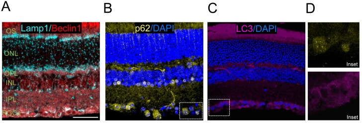
Figure 4.
Immunolabelling of autophagy proteins in eye cryosections from wild type mouse. (A) Lysosomes are immunolabeled using antibodies against Lamp1 (1D4B Developmental Sudies Hibridoma Bank, in cyan) and the autophagy regulator Beclin 1 (SantaCruz Biotechnology sc-48381, in red); (B) p62 (Progen GP62-C) is labelled in yellow and retinal nuclei are labelled with DAPI (in blue); (C) endogenous LC3 staining (Nanotools, 5F10, in magenta) with DAPI nuclear labelling (in blue). Scale bar 20 μm; and (D) insets for p62 and LC3 stainings.

