
Fig. 1. Temperature affects the formation of non-bilayer structures and ATP synthase activity in mitochondria.
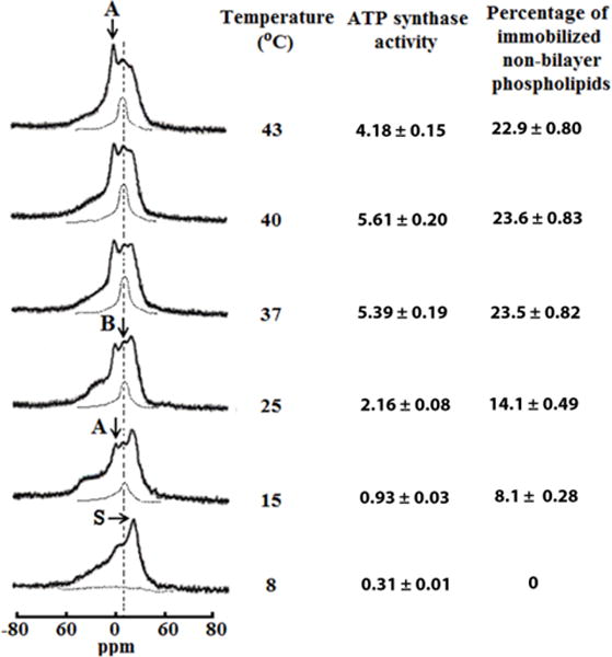
31P-NMR spectra of mitochondria recorded at various temperatures in the presence of 1.5mM succinate. The phospholipid concentration for each mitochondrial sample was estimated to be approximately 6.3 × 10⁻² M, as assessed by normalizing the integral intensity of the 31P-NMR signals from the mitochondrial samples to the integrated intensity of the 31P-NMR signals measured from large multilamellar liposomes. Hatched lines are saturation spectra observed after applying a DANTE train of saturation pulses at the high-field peak of the lamellar spectrum (see arrow with letter S). Position of the hatched line signals in saturation spectra coincides with the position of 31P-NMR signal B. The amount of non-bilayer immobilized phospholipids was estimated by calculating the area under the hatched 31P-NMR lines below the signal B after a DANTE train of saturation was applied. The percentage of non-bilayer immobilized phospholipids are shown on the column on the right as means with standard errors (±SEM) compiled from three independent experiments. Statistical differences were observed between ATP synthase activity and amount of non-bilayer phospholipids in mitochondria incubated at 25 °C vs. 15°C (One Way ANOVA, Tukey’s test). ATP levels expressed as μmol ATP synthesized per mg of mitochondrial proteins, was monitored by taking measurements on aliquots from the 31P-NMR sample tubes. ATP levels are shown on the left column as means with standard errors (±SEM) compiled from three independent experiments. Each 31P-NMR spectrum shown is representative of two independent experiments that showed similar results. Each sample was measured in triplicate readings.