
Fig. 5. CL binds avidly to DCCD-BPF and CTII.
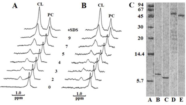
31P-NMR spectra of PC and CL in preparations containing 1.2 × 10⁻⁶ M DCCD-BPF (A) or 1. 2 × 10⁻⁶ M cardiotoxin CTII (B) in 10 mM Tris-HCl, pH 7.5, 0.5 mM EDTA, 1% Triton X-100. In the initial preparation, concentrations of PC and CL were 8.4 × 10⁻⁶ M and 4.8 × 10⁻⁶ M, respectively. Spectra were recorded at 15 °C for 30 min after adding the indicated increasing amounts of moles of exogenous CL per mol of protein. The spectra at a very top in A and B contained 1% SDS and 9 mol of exogenous CL per mol of protein. Native PAGE (C), done according to Segal et al. (1993), included standard proteins from Sigma Chemical Co., St. Louis, MO (A), DCCD-BPF (B), CTII (C), DCCD-BFP with CL (D) and CTII with CL (E). Note: for visual clarity, the lanes that are not essential for the message of this study were cropped from the same Native PAGE as indicated by white vertical lines.