Figure 1.

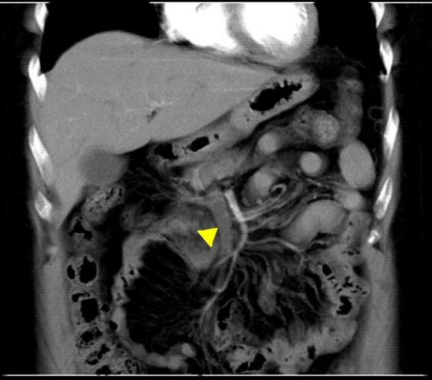
CT of the abdomen done at admission with intravenous contrast revealed thrombus in the SMV extending into the portal vein (yellow arrow head). CT: computed tomography; SMV: superior mesenteric vein.

CT of the abdomen done at admission with intravenous contrast revealed thrombus in the SMV extending into the portal vein (yellow arrow head). CT: computed tomography; SMV: superior mesenteric vein.