Fig. 7b.

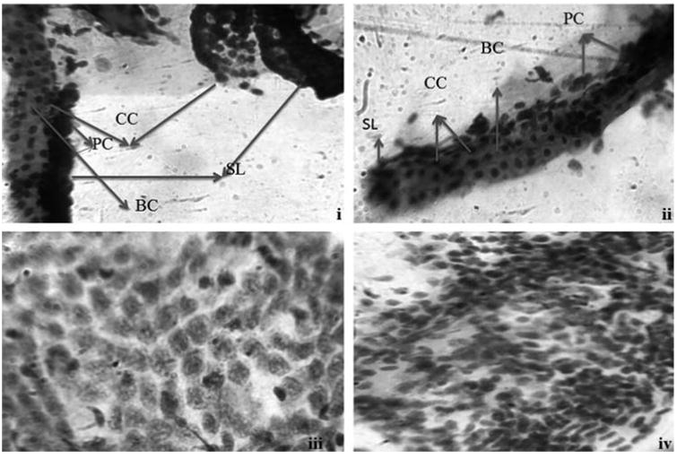
Photomicrographs of normal gills (i) and liver (iii) tissues stained with PAS staining. Photomicrographs of AgNPs exposed gills (ii) and liver tissues (iv). CC-chloride cells; PC-pavement cells; BC-blood channel; SL-secondary lamella.

Photomicrographs of normal gills (i) and liver (iii) tissues stained with PAS staining. Photomicrographs of AgNPs exposed gills (ii) and liver tissues (iv). CC-chloride cells; PC-pavement cells; BC-blood channel; SL-secondary lamella.