
Figure 4.
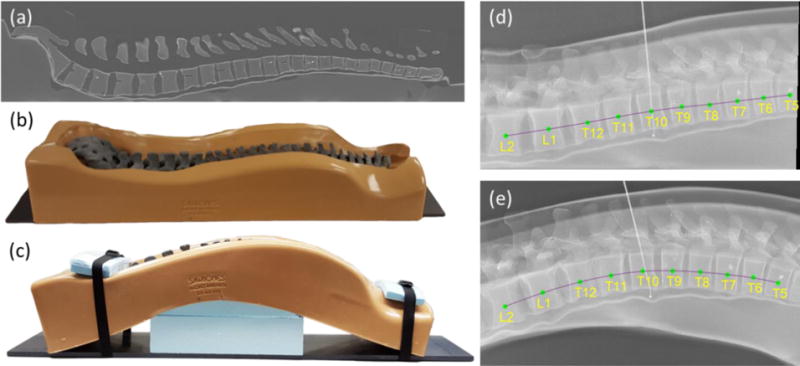
Investigation of spinal deformation in phantom. (a) Sagittal CT slice of the (b) spine phantom lying flat. (c) Photograph of the spine phantom with maximally induced curvature. (d) Lateral radiograph with vertebral levels overlaid of the phantom lying flat (as in (b)). (e) Lateral radiograph of the phantom with maximal deformation (as in (c)), overlaid with level labels.