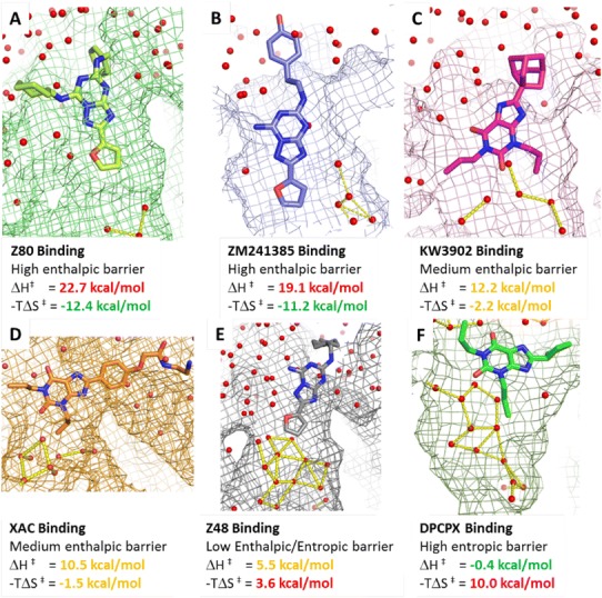
Fig. 5.

Protein–ligand locations/conformations corresponding to the binding kinetic bottlenecks detected by the suMetaD protocol for the 6 small molecules considered in this study. The ligand is shown in stick representation, the pocket as a mesh surface and waters as small spheres. Interactions among waters in the orthosteric site are shown as yellow dotted lines. The experimental energy of the enthalpic (ΔH‡) and entropic (−TΔS‡) components of the TS is reported
