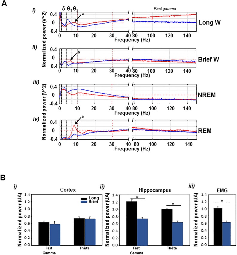
Fig 3. Spectral analysis of sleep stages normalized to the total spectrum of the signal in 24 hours.
A) Hippocampal spectral content of long and brief wake is different (in red; cortex in blue); i and ii, long wake epochs had an augmented theta band with a characteristic 8 Hz peak (θ2 band; arrow a), whereas in the brief wake a lower frequency theta peak activity was at 6 Hz (θ1 band; arrow b); in addition, the power of fast gamma hippocampal frequencies in long wake epochs is significantly increased compared to brief wake. During nrem sleep a predominance of delta and beta power, and a reduction of gamma band were observed (iii) and in rem there was a predominance of theta (iv; arrow a) with low delta and beta, and an increased gamma band with respect to nrem. B) Long vs. brief wake spectrums; hippocampal, but not cortical, fast gamma and theta power (θ2) and EMG tone were decreased in brief-wk (p < 0.001).

