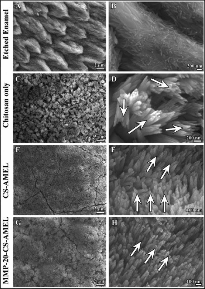
Figure 2.

Microstructure of enamel-like apatite crystals in the newly grown layer. Scanning electron micrography images showing (A, B) etched enamel, newly grown hydroxyapatite crystals in chitosan hydrogel (C, D), amelogenin-chitosan hydrogel (E, F), and amelogenin-chitosan hydrogel with matrix metalloproteinase–20 (G, H). Arrows in D, H, and F indicate the crystal orientation.
