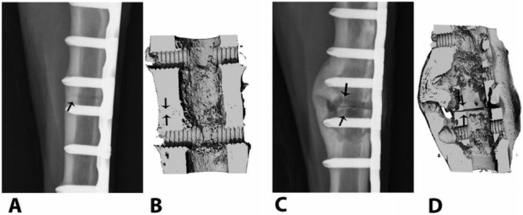
Figure 4.
Osteotomies repaired with vancomycin-tethered plates (A, B) and untreated, control plates (C, D). The representative craniocaudal digital radiograph of the site with the vancomycin-tethered plate made 90 d after osteotomy (A) is consistent with normal bone healing and callus formation at the osteotomy site (arrow), but the site with the control plate (C) shows progressive signs of cortical thinning, periosteal disruption, and osteolysis consistent with septic osteomyelitis at the osteotomy site (arrows). Three-dimensional reconstructed microcomputed tomography views of the same osteotomy sites in the vancomycin-tethered (B) and control animals (D) show persistence of the osteotomy gap (arrows) in the control, with a poorly organized lytic callus, enlarged medullary canal, and cortical thinning. Reproduced by permission from Stewart et al. (2012).

