Figure 3.
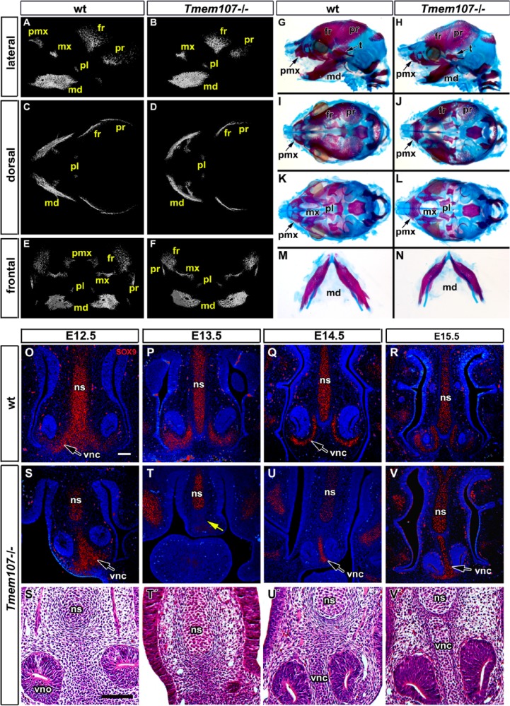
Reduced and delayed craniofacial skeletogenesis in Tmem107–/– embryos. Micro–computed tomography (CT) analysis of Tmem107–/– animals at E15.5 (A–F). Lateral view shows altered morphology and level of mineralization in maxillary, mandibular, and parietal bones in Tmem107–/– mutants and absence of the premaxillary bone (A, B). Ventral view reveals deformed mandibular bone and shorter frontal and parietal bones in Tmem107–/– embryos (D) compared to wild-type (wt) (C). On frontal view, mandibular bones have a different shape and premaxillary bone is not well mineralized in Tmem107–/– mutants (F) in contrast to wt animals (E). Alcian blue/Alizarin red staining at E16.5 demonstrates delayed mineralization and altered morphology of frontal, parietal, and premaxillary bones in Tmem107–/– embryos (H, J) in comparison to wt (G, I). Palatine bones and palatal process of the maxillary bone are reduced in Tmem107–/– mutants (L) compared to wt (K). Mandibular bones are deformed in Tmem107–/– embryos (N) in contrast to wt animals (M). SOX9 expression in developing cartilages of interorbital septum and surrounding vomeronasal organ in wt animals through the palate development (O–R). Pattern of SOX9 expression is altered in Tmem107–/– mutants in correspondence to cartilage phenotype (S–V). Cartilages surrounding the vomeronasal organ exhibit altered shape; they were fused in midline (S′, T′, U′, V′) or missing (yellow arrow in T, T′). Nuclei are counterstained by DAPI (blue). fr, frontal bone; md, mandibular bone; mx, maxillary bone; ns, nasal septum; pl, palatal bone; pmx, premaxillary bone; pr, parietal bone; t, temporal bone; vnc, vomeronasal cartilage; vno, vomeronasal organ. Scale bar = 100 µm.

