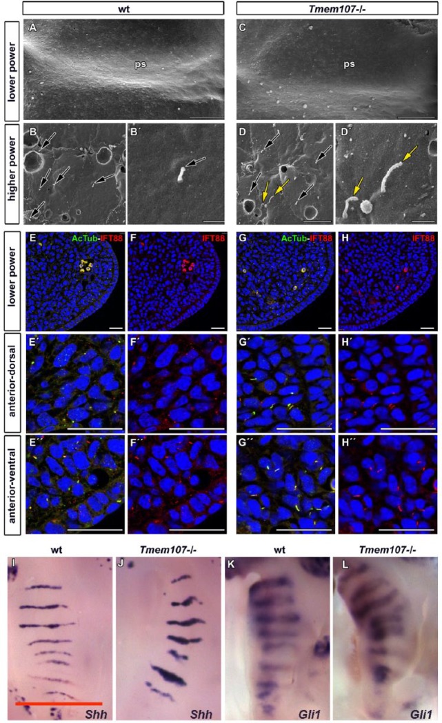
Figure 5.
Cilia morphology, IFT88 expression, and Shh pathway signaling are altered in Tmem107–/– mutants. Low-magnification scanning electron microscope images of palatal shelves (ps) at E12.5 (A, C). Higher magnification examination of anterior, middle, and posterior areas of the palatal shelve surfaces revealed altered length and morphology of primary cilia in Tmem107–/– mutants (D, D′) compared to controls (B, B′). Numerous elongated, curly cilia or bulges on cilia tip (yellow arrows) or short cilia (black arrows) were detected. Scale bars for A and E = 100 µm, scale bar for B–D and F–H = 1 µm. Primary cilia labeled by α-acetylated tubulin appear longer and have altered shape in Tmem107–/– (G′–H′′) compared to wild-type (wt) (E′, F′′) in both ventral and dorsal parts of the anterior palatal shelves. IFT88 expression is more abundant in several cilia of Tmem107–/– mutants (D) in comparison to wt animals (B). Cilia were labeled with α-acetylated tubulin (green) and IFT88 (red) in developing palatal shelves (A–D′′). Scale bar = 25 µm. Palatal views on E14.5 embryos exhibit higher expression of Shh and Gli1 in the area of palatal ridges for both analyzed genes in Tmem107–/– mice (J, L) in comparison to littermate controls (I, K) as visualized by in situ hybridization. Scale bars = 1 mm.

