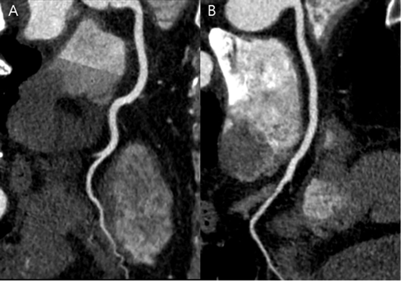
Fig 3. CT images of right coronary artery obtained in APSCM group with 100 kV and in matched BMI group.
(A) Curved multiplanar reformatted image of RCA in a 43-year-old male in the APSCM group with a BMI of 30.1 kg/m2 and heart rate of 46 beats per minute. Image was obtained in the axial mode (100 kV and 350 mAs). Mean image quality score was 4, mean contrast enhancement was 580.3 HU, mean CT number was 483.9 HU, mean image noise was 40 HU. Mean CNR and SNR were 14.5 and 12.1, respectively. CTDIvol, DLP, and effective dose were measured at 18.3 mGy, 253 mGy⋅cm, and 3.5 mSv, respectively. (B) Curved reformatted image of RCA in a 50-year-old male in the matched BMI group with a BMI of 31.6 kg/m2 and heart rate of 59 beats per minute. Image was obtained in the axial mode (120 kV and 380 mAs). Mean image quality score was 4, mean contrast enhancement was 397.1 HU, mean CT number was 324.4 HU, mean image noise was 29.1 HU. Mean CNR and SNR were 13.6 and 11.1, respectively. CTDIvol, DLP, and effective dose were measured at 27.8 mGy, 383 mGy⋅cm, and 5.4 mSv, respectively. APSCM, automatic tube potential selection with tube current modulation; BMI, body mass index; CNR, contrast-to-noise ratio; CT, computed tomography; CTDIvol, volume CT dose index; DLP, dose-length product; RCA, right coronary artery; SNR, signal-to-noise ratio.

