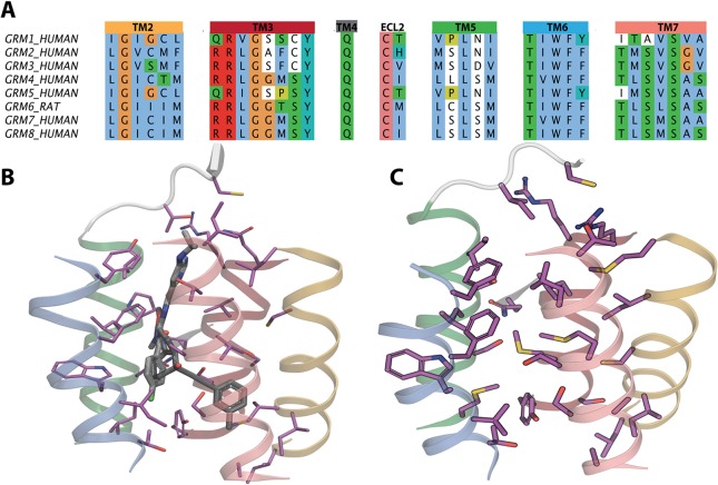
Figure 2.
(A) Nonsequential alignment of chosen binding site amino acids, coloring is based on Clustal X similarity. (B) mGlu1 and mGlu5 7-TM crystal structures showing NAMs and binding site amino acids. (C) An example of mGlu7 7-TM model receptor generated based on the sequence alignment and showing the same corresponding allosteric binding site amino acids.

