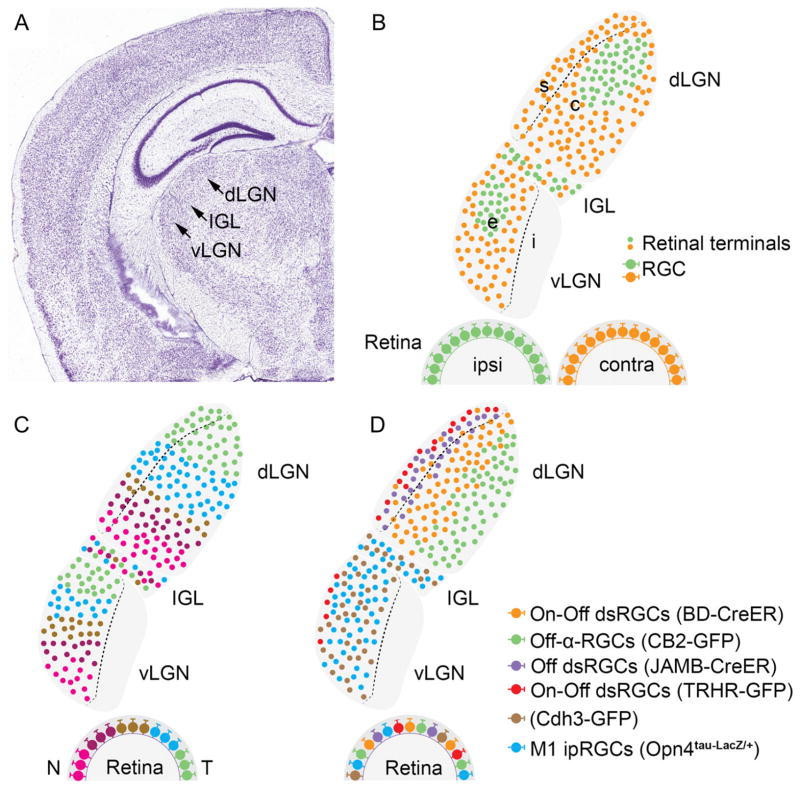
Fig. 2.
Organization of retinal projections in nuclei of the lateral geniculate complex. (A) Coronal view of a Nissl-stained mouse brain. Arrows indicate the location of dLGN, IGL, and vLGN. Image is from the Allen Brain Atlas (http://www.brain-map.org). (B–D) Schematic representation of coronal section through the lateral geniculate complex of nocturnal rodents. (B) depicts eye-specific segregation of retinal projections in dLGN, IGL, and vLGN. Terminals of ipsilateral retinal projections are depicted as green dots; terminals of contralateral retinal projections are depicted as orange dots. RGCs from which these projections arise are shown in the retinal cross sections. Dotted line in dLGN depicts the approximate boundary separating the dorsolateral shell (s) from the ventromedial core (c). The dotted line in vLGN depicts the boundary separating the external layer (e) from the internal layer (i). (C) depicts topographic mapping of retinal arbors in dLGN, vLGN, and IGL. Colors represent temporal (T) to nasal (N) location of RGCs in the retina (Feldheim et al., 1998; Pfeiffenberger et al., 2006; Huberman et al., 2008a). (D) depicts class-specific targeting of RGC axons to distinct sublamina of dLGN, vLGN, and IGL. Colors represent some classes of RGCs studied with transgenic reporter mice. Names of these reporter mouse lines are indicated in parentheses (see Hattar et al. 2006; Kim et al. 2008; Huberman et al. 2009; Kim et al. 2010; Osterhout et al. 2011; Rivlin-Etzion et al. 2011). Color-filled dots in the dLGN, IGL, and vLGN represent retinal terminals (and are not meant to indicate that these terminals innervate distinct cells). dLGN, dorsal lateral geniculate nucleus; IGL, intergeniculate leaflet; vLGN, ventral lateral geniculate nucleus; dsRGC, direction-selective retinal ganglion cell; ipRGC, intrinsically photosensitive retinal ganglion cell.

