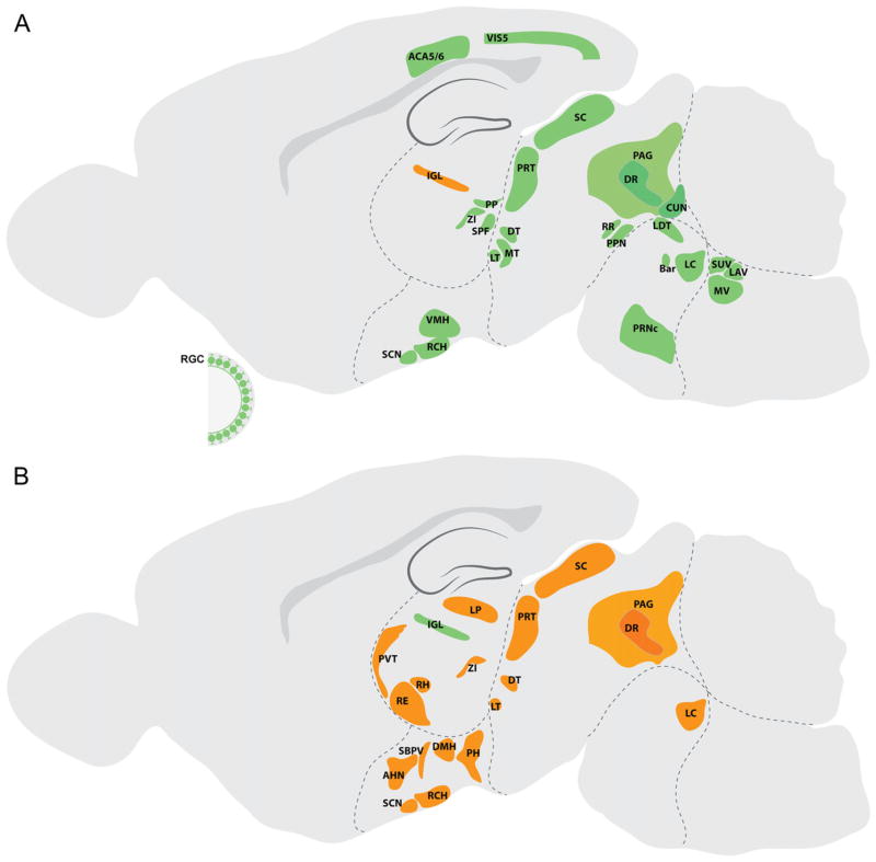
Fig. 5.
Afferent and efferent projections of rodent IGL. (A) Sources of afferent projections to IGL are colored green. (B) Brain regions innervated by IGL neurons are depicted in orange. IGL, intergeniculate leaflet; LP, lateral posterior thalamic nucleus; PP, PP; ZI, zona incerta; SPF, subparafascicular nucleus; RH, rhomboid nucleus; RE, reuniens nucleus; PVT, paraventricular nucleus; SCN, suprachiasmatic nucleus; RCH, retrochiasmatic area; SBPV, subparaventricular zone; AHN, anterior hypothalamic area; PH, posterior hypothalamic nucleus; VMH, ventromedial hypothalamic nucleus; DMH, dorsomedial nucleus of the hypothalamus; SC, superior colliculus; PRT, pretectal region; MT, medial terminal nucleus; LT, lateral terminal nucleus; DT, dorsal terminal nucleus; DR, dorsal raphe nucleus; PAG, periaqueductal gray; CUN, cuneiform nucleus; PPN, pedunculopontine nucleus; RR, retrorubral nucleus; LC, locus coeruleus; LDT, laterodorsal tegmental nucleus; Bar, Barrington’s nucleus; MV, medial vestibular nucleus; LAV, lateral vestibular nucleus; SUV, superior vestibular nucleus; PRNc, pontine reticular nucleus; VIS5, visual cortex, layer 5; ACA5/6, anterior cingulate area, layer 5 and 6; RGC, retinal ganglion cell.

