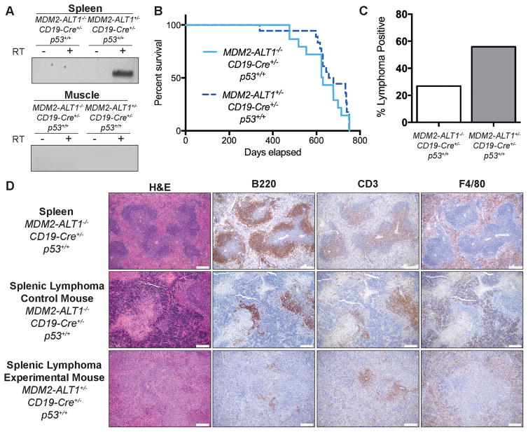
Figure 6. Mice expressing MDM2-ALT1 in B cells exhibit increased incidence of lymphomas compared to controls.
A. Expression of MDM2-ALT1 was confirmed in the spleens (B cells) of experimental mice positive for both the transgene and CD19-Cre alleles (2C12-MDM2-ALT1+/−; CD19-Cre+/−; p53+/+), but not in control mice negative for the MDM2-ALT1 transgene (MDM2-ALT1−/−; CD19-Cre+/−; p53+/+). Importantly, skeletal muscle of the transgene positive mice where the CD19 promoter is not active, did not express MDM2-ALT1. Control reactions without reverse transcription are also shown (-RT) to detect for possible contamination. B. Kaplan-Meier curves show no difference in life expectancy between survival cohorts of experimental (n = 18) and control mice (n = 15, p = 0.2737 Gehan-Breslow-Wilcoxon test). C. Mice expressing MDM2-ALT1 (19/34 lymphoma positive; 56%) in B cells showed significantly higher incidence of lymphomas compared to MDM2-ALT1 negative mice (11/41 lymphoma positive; 27%) at 18 months of age (p = 0.0174 Fisher’s exact test). D. Immunohistochemical analyses of representative splenic lymphomas from control (female – 22 months, middle panel) and experimental (female – 21 months, bottom panel) mice. For comparison, stained sections of a normal mouse spleen (MDM2-ALT1−/−; CD19-Cre+/−; p53+/+) are presented (male – 21 months, top panel). Overall, the architecture of the spleen shows marked disruption by neoplastic lymphocytes, which are more atypical in the experimental lymphoma compared to the lymphoma from the control mouse and the wild-type spleen. B220-positive B cells are significantly lowered in comparison to the normal spleen and the control mouse lymphoma and only a few small aggregates are seen scattered in the red pulp and the white pulp nodules. Additionally, CD3-positive staining is observed throughout the white pulp nodules that have lost normal architecture as compared to staining in the periarterioral lymphoid sheath in the control lymphoma and wild-type spleen. F4/80-positive cells are mainly located in the red pulp surrounding the white pulp nodules with some scattered within the white pulp; a pattern similar to normal macrophage distribution in the spleen (Scale = 200 μm).

