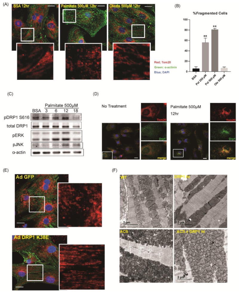
Figure 6. DRP1 mediates mitochondrial fission after lipid overload.

(A) Rat neonatal cardiomyocytes were stimulated with growth medium with or without 500μM palmitate or oleate. 12hrs after stimulation, cells were fixed with 4% paraformaldehyde and immunostained with α-actinin (green), Tom20 (red) and DAPI (blue). Scale bars indicate 20μm. (B) Quantification of mitochondrial fragmentation presented in Figure 6A. More than 100 cells were counted to determine the percentage (%) of cells with fragmented mitochondria. n=3, **: P<0.01. (C) Increased phosphorylation of DRP1 at Ser616 after palmitate treatment. NRVCs were treated in growth medium with 500μM palmitate and cell lysates were harvested at the indicated times in hr. (D) NRVCs were treated in growth medium with or without 500μM palmitate and subjected to immunohistochemistry for DRP1 (green) and Tom20 (red). Note that DRP1 is co-localized with Tom20 after palmitate treatment. Scale bars indicate 20μm. (E) NRVCs were infected with AdGFP or Ad DRP1K38E and were treated in growth medium with 500μM palmitate. NRVCs were subjected to immunohistochemistry for α-actinin (green) and Tom20 (red). See also Online Fig.VII A. Scale bars indicate 20μm. (F) Representative electron micrographs of longitudinal heart sections obtained from WT, DRP1+/-, ACStg and ACStg × DRP1+/-. DRP1 knockdown partially rescued mitochondrial morphology in ACStg hearts. See also Online Fig.VII B-E.
