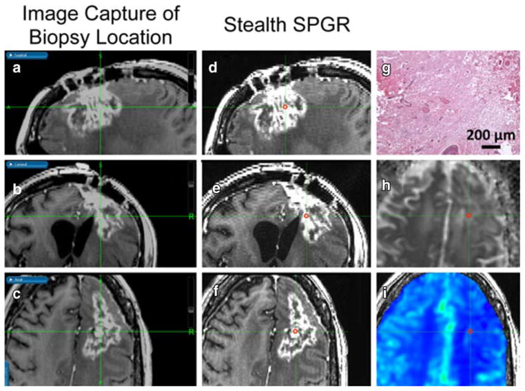
Figure 1.
3D co-localization of imaging and tissue sample shown in a 31-year-old female with TE. As outlined in red, a spherical ROI was manually drawn on the pre-surgical post-contrast Stealth SPGR (D, E, F) by visually matching it to the post-contrast Stealth SPGR images (A, B, C) captured during surgery of the tissue sample location in the sagittal (A, D), coronal (B, E), and axial (C, F) planes. Also shown in the axial plane are the corresponding ADC (H), rCBV (I), and H&E histology staining (G) showing pure TE.

