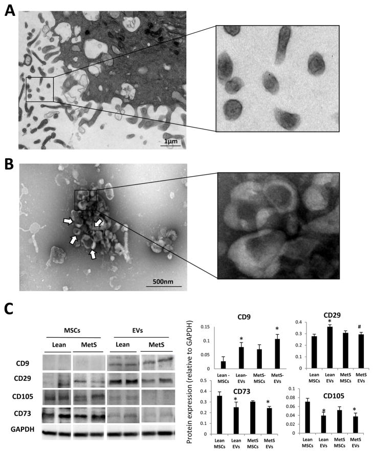
Figure 2. Characterization of MSC-derived EVs.
A: Transmission electron microscopy of culture MSCs releasing EVs. B: Negative staining of MSC culture supernatants showing EV clusters (arrows) with the classic “cup-like” morphology. C: Characterization of Lean- and MetS- EVs by protein expression of common EV (CD9 and CD29) and MSC (CD105 and CD73) markers.

