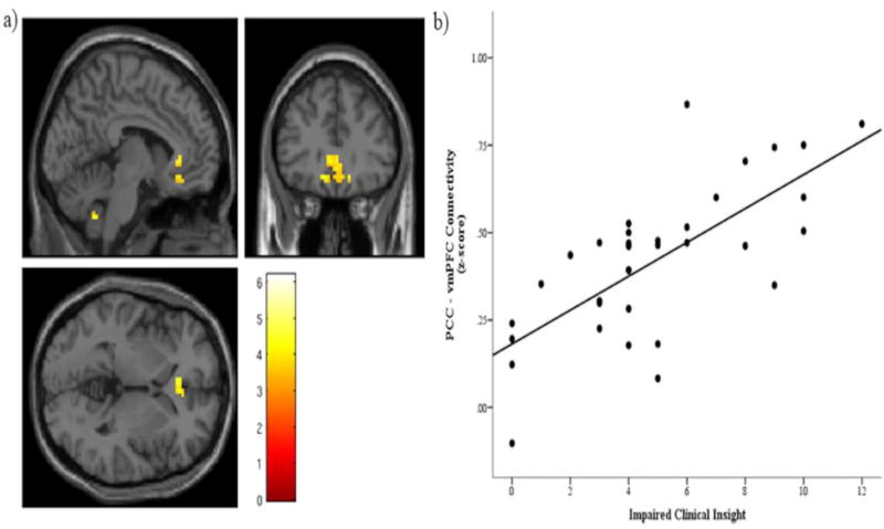
Figure 2.
Significant main effect of clinical insight predicting connectivity between the posterior cingulate cortex (PCC) and ventromedial prefrontal cortex (vmPFC). a) Significant cluster centered at MNI coordinates (−6, 30, 0), with a cluster extent of 113 voxels. A cluster forming threshold of p < .001 was applied, and this cluster was significant at the cluster level with a familywise-corrected significance of pFWE-corrected = .011. The color bar shows T values. Note, the cerebellar cluster was nonsignificant. b) Scatterplot of individual connectivity values (Fisher’s z scores) extracted from the significant cluster displayed in a), plotted against impaired clinical insight.

