
Fig. 1.
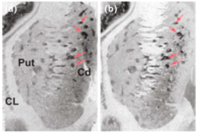
Consecutive histological sections of human striatum showing the distribution of binding sites labeled by [125I]DOI (a) and [3H]MDL100,907 (b). Arrow heads indicate some of the striosomes visualized with these radioligands. Cd, caudate; Put, putamen; CL, claustrum