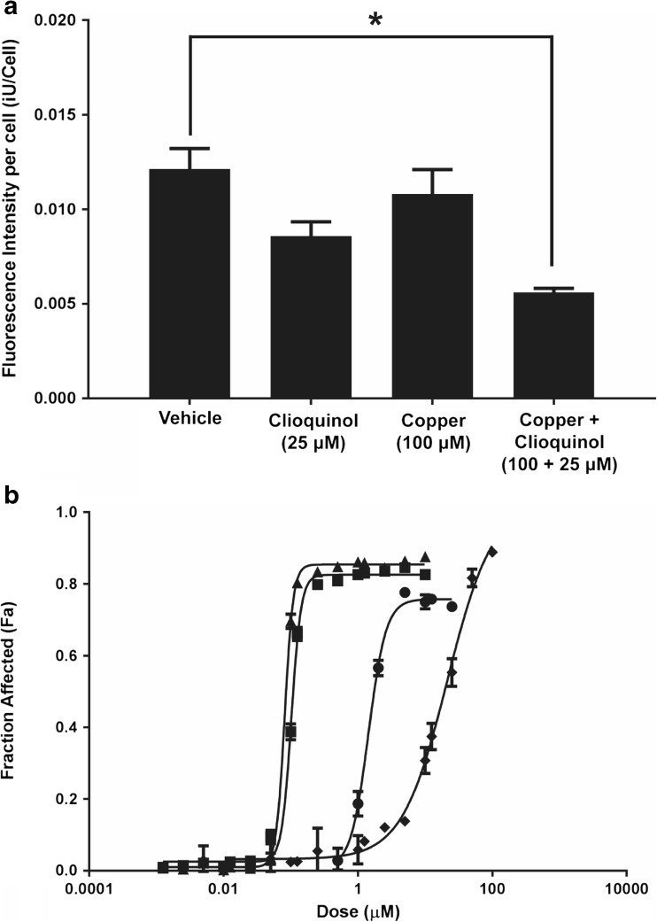
Fig. 5.
Cu(CQ)2-mediated increase in copper delivery to cells and its in vitro activity when combined with disulfiram (DSF). a A2780-CP intracellular copper levels were assessed using the cell permeable dye Phen Green™. Cell associated Phen Green™ fluorescence was measured 1-h treatment with the vehicle (0.01% DMSO), CQ, copper, or Cu(CQ)2. The cells were then incubated with Phen Green™ for 30 min (see “Materials and methods”). The fluorescence of the probe is quenched in the presence of Cu, and thus a decrease in cell associated fluorescence is indicative of higher intracellular copper levels. Cell-associated fluorescence was measured using an INCell Analyzer 2200. Results shown are an average of three studies done in triplicate (mean ± SEM). b Cytotoxicity curves were generated in A2780-CP cells after 72-h treatment with DSF (-●-), Cu(CQ)2 (-♦-), or DSF in combination with Cu(CQ)2 (-■-), or CuSO4 (-▲-). Cell viability was determined using an INCell analyzer 2200, where viability was assessed based on loss of plasma membrane integrity 72 h following treatment, i.e., total cell count and dead cell count were determined using Hoechst 33342 and ethidium homodimer staining, respectively

