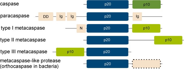
Fig. 1.
Schematic domain organisation of the C14 cysteine proteases. Domains were identified using InterPro protein sequence analysis and classification tool. The catalytic p20-like domain is coloured in dark blue and the p10 domain in green; light green indicates the presence of a 280-loop involved in calcium binding found in metacaspases. Additional domains are coloured in light red. A dashed border indicates the presence or absence of additional domains. Figure is not drawn to scale. Ig immunoglobulin-like domain, DD death domain, N N-terminal proline-rich repeat

