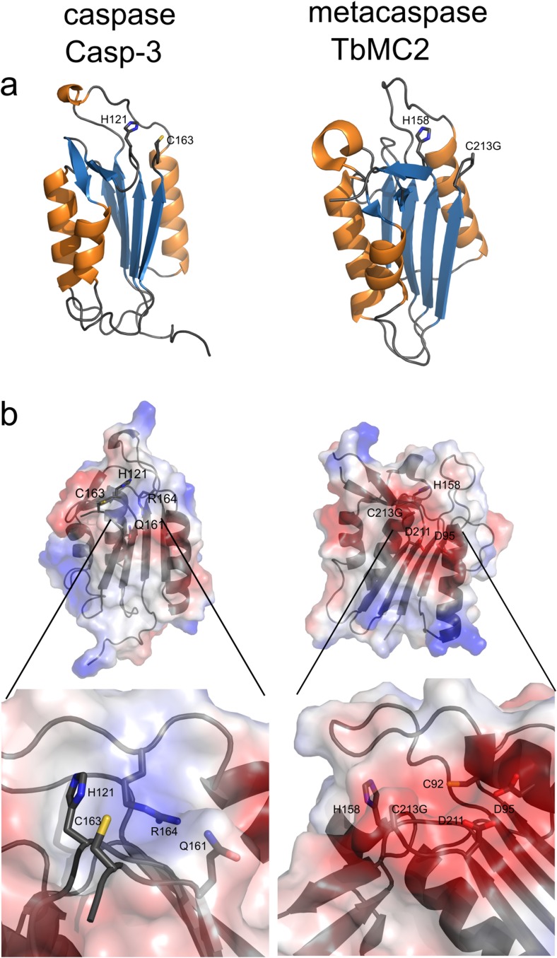
Fig. 2.
Comparison of the properties of p20-fold and specificity pocket in caspases and metacaspases. The p20 domain of caspase-3 (Casp-3), PDB ID: 3gjt (Fang et al. 2009) is compared with the type I metacaspase TbMC2, PDB ID: 4af8 (McLuskey et al. 2012). a Ribbon representation of the p20 domains: α-helices are coloured in orange and β-sheets in blue, side chains of the amino acid residues of the catalytic dyad are shown as sticks. b Surface potentials of caspase-3 and TbMC2; blue indicates basic amino acids, red acidic amino acids. The inlets display the specificity pockets in more detail. Side chains of the amino acids in the catalytic dyad and specificity pocket are shown as sticks

