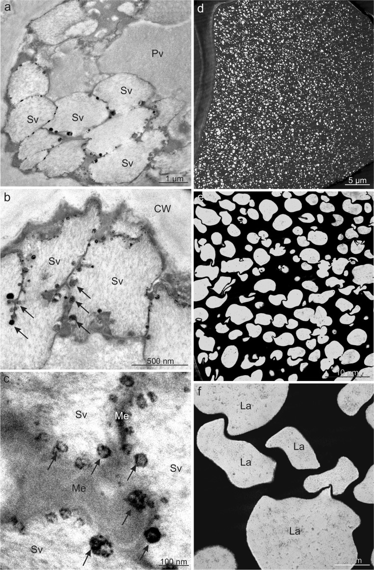
Fig. 8.
Ultrastructure of fruit epidermis cells of V. opulus (a–c) and V. lantana (d–f) in TEM. a, b Visible numerous secondary vacuoles near the cell wall with electron-dense tannin deposits (arrows) near membranes. c Tannin deposits (arrows) located around membranes of secondary vacuoles. d, e In the vacuole, electron-dense tannin complexes with numerous lacunae. f Vacuolar lacunae filled with flocculent content; CW cell wall, Pv primary vacuole, Sv secondary vacuole, Me membrane, La lacunae

