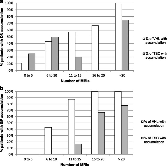
Fig. 2.

Different dynamics of Gd accumulation in dentate nucleus (DN) (a) and globus pallidus (GP) (b), in VHL and TSC groups. In both groups, the prevalence of patients with spontaneous hypersignal on unenhanced T1-w MRI in the DN and GP increased linearly with the number of Gd enhanced MRIs. The increase was highest in the VHL group
