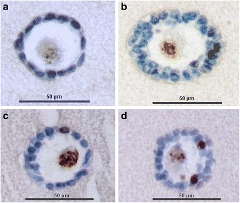
Fig. 3.

Immunohistochemistry of isolated preantral follicles embedded in a fibrin scaffold. a Embedded isolated primordial follicle with Ki67-negative granulosa cells in blue; b Embedded isolated secondary follicle with Ki67-negative granulosa cells in blue; c Embedded isolated primary follicles with one Ki67-positive granulosa cell in brown; d Embedded isolated secondary follicles with two Ki67-positive granulosa cells in brown
