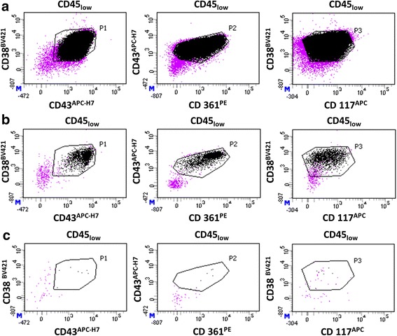
Fig. 6.

Quantification of leukemic cells by MFC in an ovarian suspension containing isolated follicles. Only viable nucleated cells (SYTO13+/7-AAD−), which are CD45low were represented (purple dots). Black dots represent leukemic cells detected with a typical immunophenotype CD38+/CD43+/CD361+/CD117+ among the population of CD45low cells. A positive event must be at the intersection of gates P1 (CD38 versus CD43), P2 (CD43 versus CD361), and P3 (CD38 versus CD117). a Positive control (leukemic cells); b MFC analysis of follicles isolated from healthy ovarian suspension after addition of 106 leukemic cells, before any wash (n = 2069 events detected); c MFC analysis of isolated follicles after 3 washes (n = 7 events detected)
