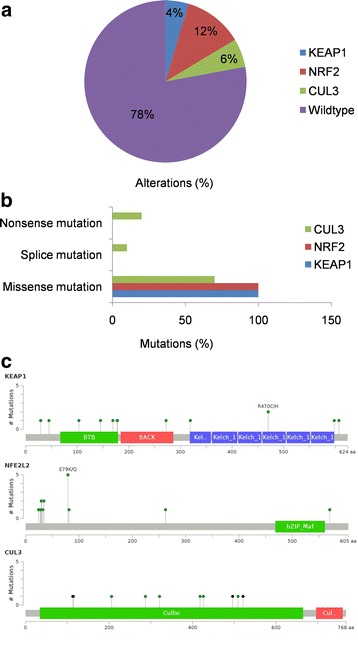
Fig. 2.

Overview of genetic changes in KEAP1-NRF2-CUL3 in TCGA-HNSCC patients. a Pie chart showing individual percentages of genetic alterations in the KEAP1-NRF2-CUL3 complex. b Bar chart showing the types and percentages of mutations of the KEAP1-NRF2-CUL3 complex. c cBioportal-predicted mutation maps (lollipop plots) showing the positions of mutations on the functional domains of KEAP1, NRF2, and CUL3 proteins. The colored lollipops show the positions of the mutations as identified by whole-exon sequencing
