Fig. 1.

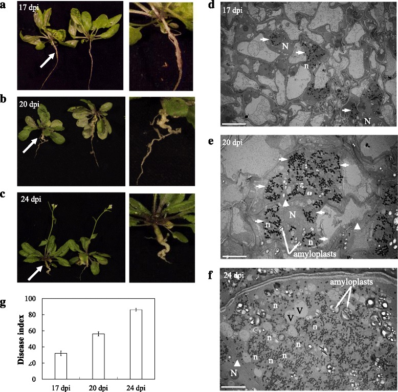
a-c Phenotype of roots in Arabidopsis plants infected by P. brassicae. a Initial symptoms of pathogen infection in roots by swelling at 17 dpi. b Formation of visible galls in roots at 20 dpi. c Deformed roots with big galls expanded through the roots at 24 dpi. d-f TEM micrographs of ultrastructural features of P. brassicae development and host cell showing magnification of the area indicated by the white arrows. d Uninucleate secondary plasmodium (white arrow) with abundant lipid droplets (black dots) in cortical cells at 17 dpi. e Multiple secondary plasmodia with abundant lipid droplets in cortical cells at 20 dpi. Thin or broken cell walls are shown by white arrowheads. f Multinucleate secondary plasmodia in the host cell at 24 dpi. Thin cell wall is shown by a white arrowhead. Amyloplasts/starch granules and small vacuoles are spotted. N: host nucleus; n: pathogen nucleus; v: vacuole. Scale bar: 10 μm. g Disease index of Arabidopsis plants at 17, 20 and 24 dpi. The data are the mean of three independent experiment. Error bars show the standard error of mean (SEM)
