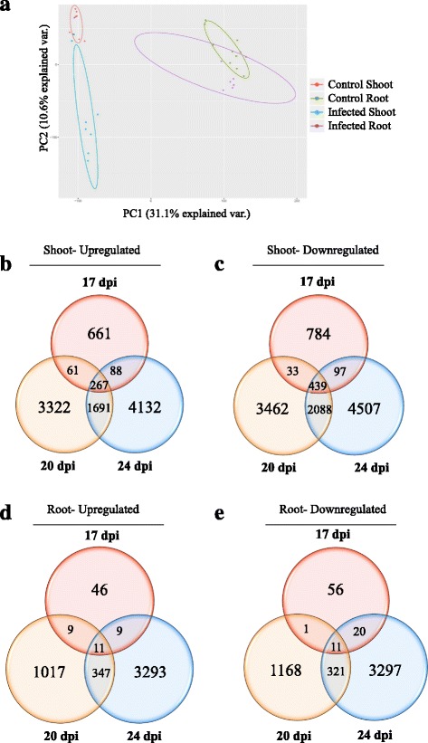
Fig. 2.

a PCA plot displaying biological variation between infected and control samples in shoot and root. b-e Venn diagrams showing the total number of significantly DEGs (p-value ≥0.05) at 17, 20 and 24 dpi in infected shoot and root compared to mock-infected control samples. The overlapping regions correspond to the number of DEGs present at more than one time point. b Up-regulated DEGs in shoot c Down-regulated DEGs in shoot. d Up-regulated DEGs in root e Down-regulated DEGs in root
