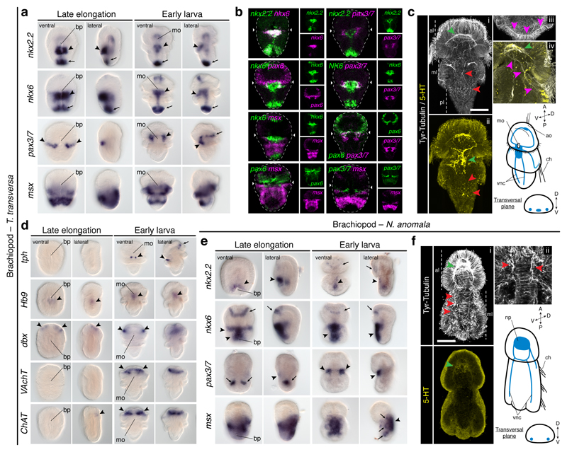
Figure 3. Dorsoventral patterning in Brachiopoda.
(a) nkx2.2 and nkx6 are in the trunk midline (arrowheads), posterior tip (arrows), gut, and apical cells (nkx6); pax3/7 is expressed laterally (arrowheads) and in the apical lobe (arrow); msx is in the mantle and ventral pedicle. (b) There is an nkx2.2+/nkx6+ medioventral region, and a more lateral nkx6+/pax6+/pax3/7+ anterior trunk domain. (c) T. transversa larval CNS (green arrowheads indicate the neuropile in i, and the trunk serotonergic condensation in ii; red arrowheads mark the VNCs; pink arrowheads indicate the innervation of the chaetae). (d) Only tph is expressed in the trunk (arrows and arrowheads indicate expression areas). (e) nkx2.2 and nkx6 are in the trunk ventral midline (arrowheads), apical lobe (arrows), and gut; pax3/7 is in the mesoderm (arrows), and in two ventrolateral trunk domains (arrowheads); msx is in the trunk, shell epithelium (arrowhead), and mesoderm (arrows). (f) N. anomala larval CNS (green arrowhead indicates the neuropile; red arrowheads in i mark the VNCs; red arrowheads in ii indicate the innervation of the chaetae). ao, apical organ; bp, blastopore; ch, chaetae; mo, mouth; np, neuropile; vnc, ventral nerve cord. Scale bars, 50 µm.

