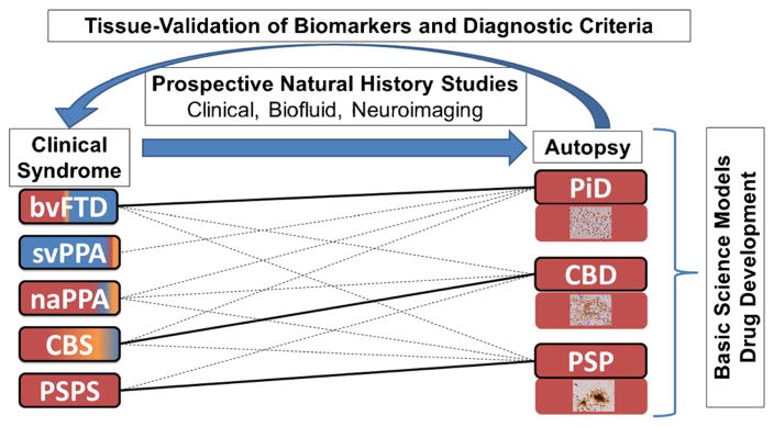
Fig. 1.
The importance of autopsy confirmation in improvement of diagnosis and treatment of tauopathies. Figure depicts clinicopathological associations of the three main FTLD-Tau neuropathologies found at autopsy with clinical syndromes. Solid lines represent the strongest associations (i.e., PiD with bvFTD, CBD with CBS, and PSP with PSPS) and dashed lines represent less frequent associations. Color shading of clinical phenotype boxes depict the relative frequencies of neuropathologies found at autopsy in each syndrome (red FTLD-Tau, blue FTLD-TDP, yellow AD) and photomicrographs in each neuropathology box depict characteristic inclusion morphologies (PiD Pick bodies, CBD astrocytic plaque, PSP tufted astrocyte). Schematic illustrates how detailed multimodal evaluations of patients with longitudinal clinical, biofluid, and neuroimaging assessments followed to autopsy can improve existing clinical criteria for detection of FTLD-Tau and differentiation from other neurodegenerative diseases and provide tissue validation for biomarkers obtained during life. Autopsy tissues also provide critical source of human-derived pathogenic tau species for use in animal/cell models of disease and therapeutic response to accelerate the development of disease modifying therapies. naPPA non-fluent agrammatic variant of primary progressive aphasia, svPPA semantic variant of primary progressive aphasia

