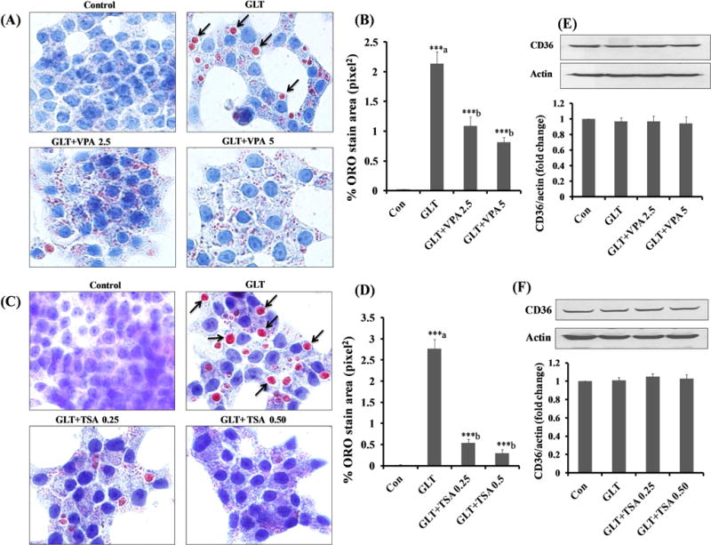
Figure 3. HDAC inhibitors markedly reduce GLT-induced lipid accumulation without affecting the CD36 expression in INS-1 832/13 cells.

Panel A–D: INS-1 832/13 cells were incubated in a medium containing low glucose (2.5 mM) with 3.75% BSA (Con), glucolipotoxicity (20 mM glucose plus 0.5 mM palmitate) in the absence and presence of VPA (2.5 or 5 mM) and TSA (0.25 and 0.50 μM) for 24 hrs. Lipid accumulation was determined by Oil-Red-O (ORO). The arrows indicate the lipid droplets accumulated under GLT and stained intense red color with ORO, whereas treatment with VPA and TSA decreased the lipids droplets. Data represent mean ± SEM of 12–15 images of each condition from three independent experiments, and are expressed as % ORO stain area in pixel2. ***p < 0.001 ‘a’ vs. con and ‘b’ vs. GLT.
Panel E and F: INS-1 832/13 cells were incubated in a medium containing low glucose (2.5 mM) with 3.75% BSA (Con), GLT (20 mM glucose plus 0.5 mM palmitate) in the absence and presence of VPA (2.5 and 5 mM) and TSA (0.25 and 0.50 μM) for 24 hrs. CD36 expression under these conditions was quantified by densitometry. Data are expressed as mean ± SEM from three independent experiments.
