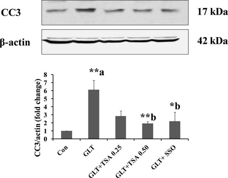
Figure 4. TSA or SSO attenuate GLT-induced caspase 3 activation in INS-1 832/13 cells.

INS-1 832/13 cells were incubated in a medium containing low glucose (2.5 mM; Con), GLT (20 mM glucose plus 0.5 mM palmitate) in the absence and presence of SSO (200 μM) and TSA (0.25 or 0.50 μM) for 24 hrs. as indicated. The cells were treated with SSO for 1 hour before the exposure to GLT. Abundance of cleaved caspase 3 (active) in cell lysates was quantified by Western blotting and densitometry. Data are expressed as mean ± SEM (n=3). **p < 0. 01 and *p < 0.05 ‘a’ vs. con and ‘b’ vs. GLT.
