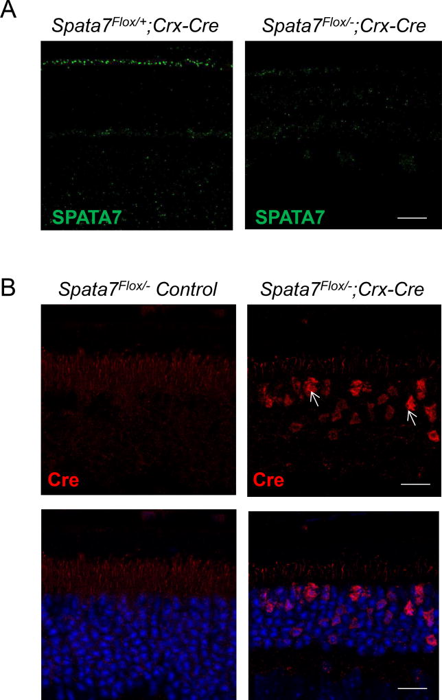
Figure 2. Loss of SPATA7 expression in the retina of Spata7Flox/−; Crx-Cre mice.
(A) Immunostaining for SPATA7 (green) in retinal sections obtained from Spata7Flox/+; Crx-Cre control mice and Spata7Flox/−; Crx-Cre mice at P28. (B) Immunostaining for Cre-recombinase in Spata7Flox/− control mouse retina and Spata7Flox/−; Crx-Cre adult cKO mouse retina at P28. Nuclei are counter-stained with DAPI (blue). Scale bar = 20 µm. CC: connecting cilium; ONL: outer nuclear layer; INL: inner nuclear layer; GCL: ganglion cell layer.

