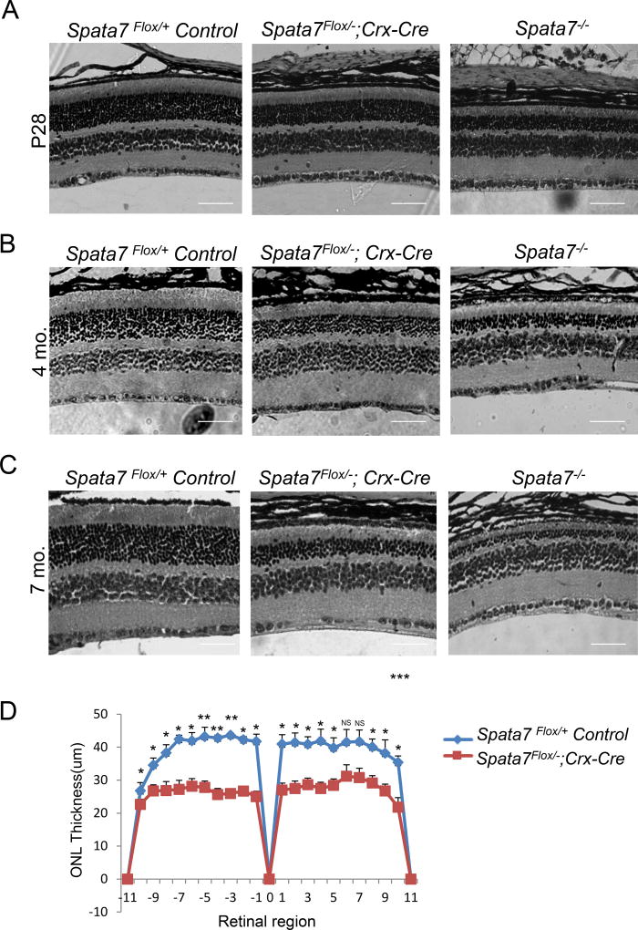
Figure 3. Progressive photoreceptor degeneration in Spata7Flox/−; Crx-Cre conditional knockout mice.
(A) Hematoxylin and eosin (H&E) staining of paraffin-embedded retinal sections obtained from Spata7Flox/+ Control, Spata7Flox/−; Crx-Cre, and Spata7−/− mice at P28 (B) 4 months, and (C) 7 months of age. Progressive thinning of the ONL is observed in Spata7Flox/−; Crx-Cre conditional KO mice and Spata7−/− (germline null) mice starting at P28, while the retina appears normal in Spata7Flox/+ control mice. (D) Retinal morphometry is presented by measuring the thickness of the ONL at 20 equally spaced positions in paraffin embedded sections along the vertical meridian of the retina for three retinas of the same genotype at P28. Three measurements were taken for each position, and a mean value was calculated. Each point represents the mean ±SEM obtained for each group (n ≥3 mouse retinas). Position 0 corresponds to the optic nerve head. Statistical analysis was performed using the Student’s t-test (NS= not significant, p>0.05; *p<0.05; **p<0.01). Scale bar = 40 µm. ONL: outer nuclear layer; INL: inner nuclear layer; GCL: ganglion cell layer.

